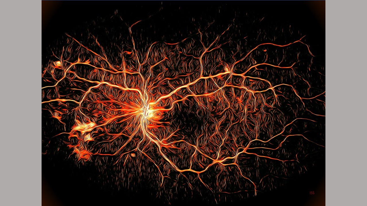
In mice and human cell models, the drug 32-134D lowered protein levels known to contribute to diabetic macular edema and diabetic retinopathy.

The research study discussed in this article was supported by funding obtained from the ICTR.
Researchers at Wilmer Eye Institute, Johns Hopkins Medicine say they have evidence that an experimental drug may prevent or slow vision loss in people with diabetes. The results are from a study that used mouse as well as human retinal organoids and eye cell lines. Eye conditions that cause vision loss are common complications of diabetes, affecting nearly 8 million Americans — a statistic likely to almost double by 2040, according to the National Institutes of Health.
The team focused on models of two common diabetic eye conditions: proliferative diabetic retinopathy and diabetic macular edema, both of which affect the retina, the light-sensing tissue at the back of the eye that also transmits vision signals to the brain. In proliferative diabetic retinopathy, new blood vessels overgrow on the retina’s surface, causing bleeding or retinal detachments and profound vision loss. In diabetic macular edema, blood vessels in the eye leak fluid, leading to swelling of the central retina, damaging the retinal cells responsible for central vision.
Results of the study, published May 25 in the Journal of Clinical Investigation, show that a compound called 32-134D, previously shown to slow liver tumor growth in mice, prevented diabetic retinal vascular disease by decreasing levels of a protein called HIF, or hypoxia-inducible factor. Doses of 32-134D also appeared to be safer than another treatment that also targets HIF and is under investigation to treat diabetic eye disease.
Current treatment for both proliferative diabetic retinopathy and diabetic macular edema includes eye injections with anti-vascular endothelial growth factor (anti-VEGF) therapies. Anti-VEGF therapies can halt the growth and leakiness of blood vessels in the retina in patients with diabetes. However, these treatments aren’t effective for many patients, and may cause side effects with prolonged use, such as increased internal eye pressure or eye tissue damage.
Akrit Sodhi, M.D., Ph.D., an author of the new study, says that in general, the idea of inhibiting HIF, a fundamental protein in the body, has raised concerns about toxicity to many tissues and organs. But when his team screened a library of HIF inhibitor drugs and conducted extensive testing, “We came to find that the drug examined in this study, 32-134D, was remarkably well tolerated in the eyes and effectively reduced HIF levels in diseased eyes,” says Sodhi, associate professor of ophthalmology and the Branna and Irving Sisenwein Professor of Ophthalmology at the Johns Hopkins University School of Medicine and the Wilmer Eye Institute.
HIF, a type of protein known as a transcription factor, has the ability to switch certain genes, including vascular endothelial growth factor (VEGF), on or off throughout the body. In the eye, elevated levels of HIF cause genes like VEGF to increase blood vessel production and leakiness in the retina, contributing to vision loss.
To test 32-134D, researchers dosed multiple types of human retinal cell lines associated with the expression of proteins that promote blood vessel production and leakiness. When they measured genes regulated by HIF in cells treated with 32-134D, they found that their expression had returned to near-normal levels, which is enough to halt new blood vessel creation and maintain blood vessels’ structural integrity.
Researchers also tested 32-134D in two different adult mouse models of diabetic eye disease. In both models, injections were administered into the eye. Five days post-injection, the researchers observed diminished levels of HIF, and also saw that the drug effectively inhibited the creation of new blood vessels or blocked vessel leakage, therefore slowing progression of the animals’ eye disease. Sodhi and his team said they also were surprised to find that 32-134D lasted in the retina at active levels for about 12 days following a single injection without causing retinal cell death or tissue wasting.
“This paper highlights how inhibiting HIF with 32-134D is not just a potentially effective therapeutic approach, but a safe one, too,” says Sodhi. “People facing diabetic eye disease and vision loss include our family members, friends, co-workers — this is a disease that impacts a large group of people. Having safer therapies is critical for this growing population of patients.”
Sodhi says that further studies in animal models are needed before moving to clinical trials.
Additional authors involved in this study are Jing Zhang, Deepti Sharma, Aumreetam Dinabandhu, Jaron Sanchez, Brooks Puchner Applewhite, Kathleen Jee, Monika Deshpande, Ming-Wen Hu, Chuanyu Guo, Jiang Qian, Shaima Salman, Yousang Hwand and Gregg Semenza of the Johns Hopkins University School of Medicine; Miguel Flores-Bellver and Maria Valeria Canto-Soler of University of Colorado School of Medicine; Nicole Anders and Michelle Rudek of the Johns Hopkins Sidney Kimmel Comprehensive Cancer Center; and Silvia Montaner of the University of Maryland.
Funding for this work was supported by NIH grants (R01EY029750, R01EY032104, EY001765, P30 CA006973, 1S10RR026824, UL1 TR003098); the TEDCO Maryland Innovation Initiative, the Research to Prevent Blindness, Inc. Special Scholar Award; the Sybil B. Harrington Stein Innovations Award; an unrestricted grant to the Wilmer Eye Institute, Johns Hopkins University School of Medicine and the University of Colorado; the Armstrong Family Foundation; the CellSight Development Fund; the Doni Solich Family Chair in Ocular Stem Cell Research; and the Branna Irving Sisenwein Professorship in Ophthalmology.
Media Contacts
